Facilitating the Collaborative Spirit of Research
Petar Grozdanov, Ph.D. and the Molecular Biology and Imaging Analysis Core Facilities transform health care through innovative technology and collaborative spirit
The new vision at Texas Tech University Health Sciences Center (TTUHSC) aims to transform health care through innovation and collaboration. Our vision is not only about the biggest names in science and leadership—everyone plays a vital role. This series seeks to highlight innovative individuals and groups that work together to create transformative ideas and shape health care, revealing what makes this university extraordinary.
The Image Analysis and Molecular Biology Core Facilities, or “the Core" facilities, are research laboratories located on the 4th and 5th floors at the TTUHSC campus in Lubbock. These facilities provide unique opportunities and support for the faculty, staff and students at Texas Tech University Health Sciences Center (TTUHSC) in their pursuit of groundbreaking research. By providing instruments that otherwise would not be available, the Core Facilities support the TTUHSC community in their drive to advance knowledge through innovative research.

“Our facilities provide state-of-the-art instrumentation to support experimental demands
for faculty, students and staff.” said Petar Grozdanov, Ph.D., director of the Image
Analysis and Molecular Biology Core Facilities. “Expertise and advice are available
for the design of experiments using microscopic and molecular biology approaches and
technologies.”
Douglas Stocco, Ph.D., founded the facilities in 2011, when he was the TTUHSC’s Executive Vice President for Research. Stocco has since retired, but the Core Facilities remain a vital part of the research process for many at TTUHSC.
“About half of my time is devoted to running the Cores, and the other half is devoted to performing my own scientific research in the Department of Cell Biology and Biochemistry in the School of Medicine,” said Grozdanov, who is no stranger to handling many tasks simultaneously. Running the Cores entails theoretical and practical application of the equipment, coordinating service and maintenance for the instruments, promoting the use of the facilities, providing training for individuals who use the instruments and overseeing the operating budget.

While Grozdanov’s personal dedication is clear and admirable, collaboration is the crucial part of what the facilities are designed to provide. Grozdanov commends and encourages that element of the research process.
“The nature of all research is collaborative,” Grozdanov said. “Nowadays, a single
laboratory or facility cannot complete all the tasks required for successful manuscript
publications or grant applications. The Core facilities encourage that collaborative
spirit.”
“Faculty members often need to provide detailed descriptions of the instruments available to them for successful grant applications on the national level,” said Grozdanov. “The Core facilities provide support to the grant applicants, enhancing their probability of obtaining funding.”

While so many elements of education have been interrupted by COVID-19, the functionality
of this space remains.
“Overall the operation of the Core facilities has stayed the same during this unprecedented time,” said Grozdanov. “This shows the resilience of the research faculty and their strong commitment to the research mission of the institution.”
Not only are these facilities safe, functional and useful for researchers, they are constantly working on expanding and improving.
“I’m always pleased to hear all the excellent feedback about issues that might arise from the use of the instruments,” said Grozdanov. “I try to emphasize how important it is to have immediate feedback if something goes wrong or does not go as expected with the instruments. In those cases, I can take appropriate action to remedy the issue and provide continuous operational service.”
In addition to Grozdanov’s commitment to the continued functionality of the laboratories’ current instruments, he also helps seek new technology for the laboratories by providing advice to the Senior Vice President for Research Min Kang, Ph.D., and the Research Council regarding the acquisition of new equipment and upgrades to existing equipment.
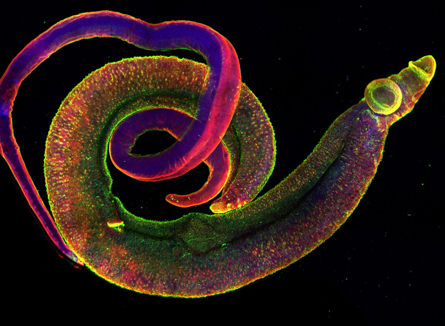

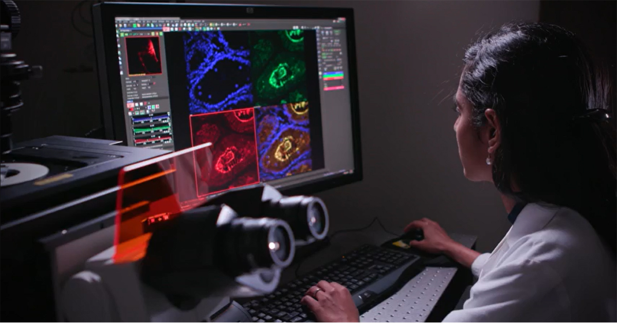
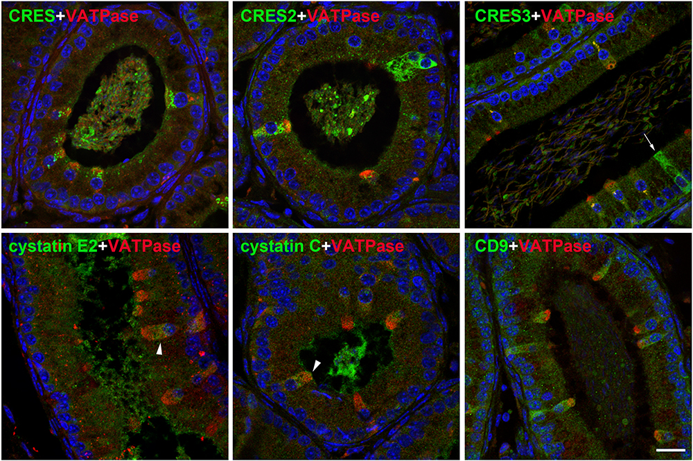
The Image Analysis and Molecular Biology Core Facilities are packed with unique, cutting-edge technology and instrumentation. When asked to highlight a particular piece of equipment, Grozdanov pointed out the confocal microscope—noting not only its scientific value, but the beauty that the imaging provides.
“My favorite instrument is the confocal microscope,” Grozdanov said, clarifying that confocal microscopy is a method to examine the structure of small samples like cells. He went on to explain that investigators use this microscope to view thin “slices” of biological samples, stacking all the slices together to get a three-dimensional view of the whole sample. An invaluable tool to TTUHSC’s scientists, it is the most frequently used of the facilities’ instruments.
“I like this microscope because the images taken can be seen as a form of art as well,” Grozdanov said. “The images display vibrant colors and shapes often never seen before, reflecting the structural and functional fabric of life.”
No matter what the research entails, any project that goes on within the Core facilities is made possible by these remarkable tools. Grozdanov takes pleasure in being available for those embarking on such incredible pursuits, and being able to assist them on their journey.
“I am proud of all the scientific discoveries that are made using the Core equipment,” said Grozdanov.
Related Stories
38th Student Research Week Successful at Showcasing Secrets of Immune Defense
Student researchers at TTUHSC had the opportunity to showcase their presentation skills at the 38th Annual Student Research Week Feb. 25 – 27.
Celebrating Veterans: TTUHSC’s General Martin Clay’s Legacy of Service and Leadership
From his initial enlistment in the Army National Guard 36 years ago to his leadership in military and civilian health care management roles, Major General Martin Clay’s career has been shaped by adaptability, mission focus and service to others.
Texas Tech University Health Sciences Center School of Nursing Named Best Accelerated Bachelor of Science in Nursing Program in Texas
The TTUHSC School of Nursing Accelerated Bachelor of Science in Nursing (BSN) program has been ranked the No. 1 accelerated nursing program in Texas by RegisteredNursing.org.
Recent Stories
Stay Safe: Preventing Summertime Dehydration
As the summer heat continues to intensify, so does the risk for dehydration. Prioritizing hydration this summer can protect you and your loved ones from potentially life-threatening complications.
Julia Jones Matthews School of Population and Public Health Offers New Graduate Certificate in Epidemiology
The TTUHSC Julia Jones Matthews School of Population and Public Health is launching a new MPH concentration in Epidemiology and a Graduate Certificate in Epidemiology.
‘Tropical Neglected Disease’ Not Forgotten by TTUHSC Researcher
Afzal Siddiqui, Ph.D., director of the TTUHSC Center for Tropical Medicine and Infectious Diseases, developed SchistoShield®, a vaccine to treat schistosomiasis, as a humanitarian effort, rather than making it for profit.