Ultrasound Is Changing How Physicians Diagnose at Patient’s Bedside
TTUHSC Hosted Fourth World Congress on Ultrasound in Medical Education

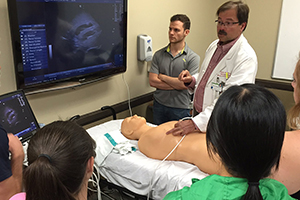
School of Medicine students use ultrasound during an anatomy review.
In the 21st century management of athletic injuries, it is likely a physician from the Texas Tech University Health Sciences Center (TTUHSC) Sports Medicine fellowship will perform a musculoskeletal ultrasonography on the player, to check for any muscle, ligament or bone injury.
Ultrasound is a high frequency sound, too high for humans to hear. Medical ultrasound or ultrasonography use these high frequency sound waves to create images of organs and structures inside the body. The names of procedures and ultrasound images change according to the specific area of the body focused on.
After a special gel, which helps ultrasound travel to the body is put on the skin, physicians, ultrasonographers or health care professionals place a transducer, an instrument sending out ultrasound to examine patients. When the sound waves meet structures inside of the body, some of them bounce off and return to the transducer as echoes. The collected echoes are processed by a computer to make images and they are displayed on a monitor.
Physicians and health care professionals use the medical ultrasonography to view anatomy or normal structure and various diseases in many different organs, measure the speed of blood flow in the heart and blood vessels and evaluate movements of heart valves or organs in the body.
As ultrasound does not use x-rays or ionizing radiation, the very first imaging study you and your children will experience in life is fetal ultrasound, nonintrusive and painless ultrasonography of a baby in the womb. The fetal ultrasonography is used to monitor the baby’s growth and health. Some babies have more than a dozen of ultrasonography if their heads are too big or they are positioned awkwardly in their mom’s uterus. Gynecologic ultrasonography is the imaging of the female reproductive system.
In addition to diagnosis, the ultrasonography has been used for treatment and guidance during procedures that require intervention such as placement of catheter into vessels, injections of drugs to joints and biopsies.
Jongyeol Kim, M.D., associate professor of the TTUHSC Department of Neurology and neurologist at Texas Tech Physicians, said several medical schools including TTUHSC School of Medicine are teaching ultrasound to medical students as regular curriculum.
“During anatomy class, the first year medical students are scanning standardized patients or live models to see organs after cadaver dissection,” Kim said. “Seeing organs and blood flow running in live human body with non-intrusive ultrasound augments the students’ learning experience when they learn anatomy, physiology and physical examination skills.”

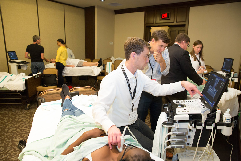
World Congress attendees perform a hands-on ultrasound workshop.
With the advancements in ultrasound, health care professionals from around the world attended the Fourth World Congress on Ultrasound in Medical Education September 23 – 25 in Lubbock. TTUHSC hosted the World Congress Conference that featured physicians, educational researchers and other experts in the field of ultrasound.
More than 400 health care professionals and students attended the conference. One conference attendee said, “What a gem of a place and such a great congress. I never would have known if I had not come.”
Ultrasonography was widely used in radiology, cardiology and obstetrics and gynecology over the past several decades, but clinicians from other specialties had not used them frequently until recently when more compact and affordable ultrasound machines were developed.
“With further advancement of technology, the ultrasound machines become more compact, portable and less expensive with higher imaging quality,” Kim said. “The availability of portable ultrasound devices enables physicians of other specialties such as emergency medicine, critical care and internal medicine to embrace point-of-care ultrasonography — ultrasonography performed and interpreted by the clinician at the bedside.”
Related Stories
Celebrating Veterans: TTUHSC’s General Martin Clay’s Legacy of Service and Leadership
From his initial enlistment in the Army National Guard 36 years ago to his leadership in military and civilian health care management roles, Major General Martin Clay’s career has been shaped by adaptability, mission focus and service to others.
Texas Tech University Health Sciences Center School of Nursing Named Best Accelerated Bachelor of Science in Nursing Program in Texas
The TTUHSC School of Nursing Accelerated Bachelor of Science in Nursing (BSN) program has been ranked the No. 1 accelerated nursing program in Texas by RegisteredNursing.org.
TTUHSC Names New Regional Dean for the School of Nursing
Louise Rice, DNP, RN, has been named regional dean of the TTUHSC School of Nursing on the Amarillo campus.
Recent Stories
The John Wayne Cancer Foundation Surgical Oncology Fellowship Program at Texas Tech University Health Sciences Center Announced
TTUHSC is collaborating with the John Wayne Cancer Foundation and has established the Big Cure Endowment, which supports the university’s efforts to reduce cancer incidence and increase survivability of people in rural and underserved areas.
TTUHSC Receives $1 Million Gift from Amarillo National Bank to Expand and Enhance Pediatric Care in the Panhandle
TTUHSC School of Medicine leaders accepted a $1 million philanthropic gift from Amarillo National Bank on Tuesday (Feb. 10), marking a transformational investment in pediatric care for the Texas Panhandle.
Texas Tech University Health Sciences Center Permian Basin Announces Pediatric Residency Program Gift
TTUHSC Permian Basin, along with the Permian Strategic Partnership and the Scharbauer Foundation, Feb. 5 announced a gift that will fund a new pediatric residency.
